e martë, 12 qershor 2007

An Overview

Tests:

Lab Tests:

  1. Amylase (the pancreatic enzyme responsible for digesting carbohydrates) is the most common blood test for acute pancreatitis. It increases from 2 to 12 hours after the beginning of symptoms and peaks at 12 to 72 hours. It may rise to 5 to 10 times the normal level and will usually return to normal within a week. Amylase also may be monitored with chronic pancreatitis, it will often be moderately elevated until the cells that produce it are destroyed.
  2. Lipase (the enzyme that, along with bile from the liver, digests fats) increases in the blood within 4 to 8 hours of the beginning of an acute attack and peaks at 24 hours. It may rise to several times its normal level and remains elevated longer than amylase. As cells are destroyed with chronic pancreatitis and as lipase production drops to less than 10% of the normal level, steatorrhea (fatty, foul-smelling stools) will form.
  3. Trypsin (which digests proteins) is thought to be the most sensitive blood test for acute pancreatitis but is not widely available and not routinely used. Stool trypsin tests are used to check for pancreatic insufficiency and may be part of a work-up for chronic pancreatitis. Trypsinogen (Immunoreactive trypsin) is a blood test that may be used to check pancreatic sufficiency in those with chronic pancreatitis.

Other lab tests that may be used to check for complications of acute pancreatitis include:

  1. Complete Blood Count (CBC) (including white blood cell count)
  2. Comprehensive Metabolic Panel (Bilirubin, liver function tests)
  3. Glucose
  4. Calcium
  5. Magnesium
  6. C-Reactive Protein
Other lab tests that may be used to check for chronic pancreatitis include:

  1. Fecal fat
  2. Trypsin
  3. Trypsinogen (Immunoreactive trypsin)

Non-laboratory tests may include:

  1. Abdominal ultrasound
  2. ERCP (endoscopic retrograde cholangiopancreatography), a flexible scope used to see and sometimes remove gallstones
  3. EUS Endoscopic Ultrasound
  4. ERG (Upper EndoscopyEsophagogastroduodenoscopy)
  5. CT (computed tomography) scan
  6. Secretin testing (not widely available) in which a tube is positioned in the duodenum to collect pancreatic secretions stimulated by IV secretin
  7. MRI (magnetic resonance imaging)
  8. MRCP
  9. Percutaneous Transhepatic Cholangiography (PTC)
  10. Wireless Capsule Endoscopy - "The Camera in a Pill"
  11. Barium Exrays
  12. Ultrasound
  13. Nuclear Scans
  14. Hemoccult Card Test

To view information on another disease, click on SOD and Pancreatitis Library.!

SOD and Pancreatitis Library


..

ERCP and PTC

ERCP (Endoscopic Retrograde Cholangiopancreatography)
..
Your doctor has decided that you need a test called Endoscopic Retrograde Cholangiopancreatography, or ERCP for short. This test is designed to image and treat disorders of the liver, bile ducts, and pancreas.
In many cases, a problem found during ERCP can be treated at the same time, thus avoiding major surgery. The procedure is usually performed by a Gastroenterologist, a doctor with special training in this field. The procedure might be a sphincterotomy, a removal of a stone, or a stent placement in order to keep a duct open.
..
How Your Liver, gallbladder, and pancreas Work
To better understand ERCP, it is important that you understand a little about how the liver, gallbladder, and pancreas work. Your liver makes a substance called bile, a bitter yellowish-green fluid which helps digest your food. A duct is a hollow tube that carries fluid from one place to another, like a water pipe in your home. Bile is collected from the liver by a series of ducts that resemble branches of a tree. These branches come together at the main "trunk" called the common bile duct which drains into the intestine. Between meals about a quarter cup of bile is stored and concentrated in a side branch of the common bile duct called the gallbladder, a small pear-shaped hollow sac nestled beneath the liver in the right upper abdomen. The pancreas usually shares a common drain with the liver via its pancreatic duct. With each meal, bile enters the small intestine where it breaks up large globs of fat into smaller globs, a first step in digestion.
A healthy gallbladder keeps bile flow moving like a mountain stream. However, when the gallbladder becomes diseased, the flow slows and the river turns into a swamp. The stagnant bile gradually crystallizes and the crystals clump together to form stones - like snowflakes making a snowball. (This is a totally separate disease from kidney stones that form in the urinary system. Having one kind of stone does not increase your risk of developing the other.)
..
Sometimes, a stone, scar tissue, or a tumor can block the ducts which drain the liver and/or pancreas. This is often an indication for ERCP which allows the most accurate means of visualizing the ducts and evaluating the problem.
..
How is ERCP performed?
ERCP is performed in the X-ray department. There is very little preparation except fasting. After IV sedation with medications to make you sleepy and very relaxed, a small, thin tube with a videocamera (endoscope) is passed through the mouth. It is carefully guided down the foodpipe, past the stomach, and into the first part of the small intestine - called the duodenum.
..
Using the special ERCP videoscope, your doctor can locate the small nipple-like opening called the ampulla which serves as the "drain" for your liver and pancreas.


A small catheter is then placed through this opening and dye is carefully injected so that X-rays can be taken. If a problem is identified during ERCP, treatment can often be undertaken at the same time. Stones in the bile duct can be removed, narrow strictures can be dilated, and tumors can be biopsied and bypassed with a stent to relieve obstruction. The test takes 30 to 90 minutes to perform and is usually painless to the patient who is sedated.
..
What Are The Risks of ERCP?
No test is 100% safe or without risk. Fortunately, ERCP can be performed in most patients without serious complications. On average, less than 10% of patients have complications from ERCP. The death rate is about 1 in 250, but these are usually sick hospitalized patients with multiple medical problems. The potential problems can be broken down into those related to the sedation, the scope procedure itself, and those specific to ERCP. Sedation risks To make the test comfortable, you will be given IV medications that make you sleepy. If you become too sleepy, it could suppress your breathing which could result in respiratory arrest. However, by monitoring your heart, blood oxygen level, and breathing closely, this can be prevented and this complication is rare.
..
Scope Risks
The thin, flexible videoscope is passed carefully and is usually well tolerated. However, in rare cases, damage to the lining of the upper digestive system can occur. This could cause bleeding or a hole in the intestinal wall. These complications might require surgery. Such damage occurs in less than 1 per 2000 cases. Another risk is choking on stomach contents during scope insertion. That is why it is so important not to eat or drink for the 8 hours before your examination.
..
ERCP Specific Risks
This test has some specific complications associated with injecting and instrumentation of the pancreatic duct. The most frequent complication by far is that of pancreatitis. In about 3 to 5% of cases, the pancreas gland becomes inflamed after ERCP and releases potent digestive juices directly into the surrounding tissue causing pancreatitis. Symptoms of pancreatitis include abdominal and back pain, nausea, and vomiting. In most cases, this complication gets better within 48 hours, but can be more severe in less than 1% of cases. This could result in a prolonged hospitalization, diabetes, fat malabsorption, and in rare cases, death. If the bile duct must be cut to relieve an obstruction or remove a stone, bleeding and perforation of the bowel may occur in 1 to 3% of cases. Failed ERCPIn some cases, the anatomy of the opening can prevent passage of the catheter into the ducts. Sometimes this is due to an intestinal pocket, or diverticulum, which distorts the normal anatomy. Failed ERCP occurs in about 3 to 5% of cases.
..
Preparing For ERCP


  1. Most ERCP exams are done in the morning. To prepare, you should not eat or drink anything after midnight the night before your test. If your ERCP is scheduled for the afternoon, you must remain totally fasting for at least 8 hours before your test.
  2. Be sure to tell your doctor about all allergies, especially those to shellfish, iodine, IVP dye, and X-ray contrast dye.
  3. Your regular medications can be taken with a sip of water on the morning of your exam - EXCEPT diabetic medications. If your are diabetic, your doctor will give you special instructions.
  4. You should be off all blood-thinning medications at least 3 days prior to your exam.
  5. If you have heart problems and have been told that you need antibiotics before procedures, alert your doctor.
  6. Please arrange for a driver to bring you home after the exam, preferably be a family member.
..
After ERCP
Usually the doctor will discuss the results right after the exam. Occasionally, an office visit will be arranged to discuss more complicated problems, or if you are too sleepy. If your test was done as an outpatient, you will usually be allowed to return home about two hours after your exam. This allows time for most of the sedation to wear off and to screen for complications. Plan to go straight home - not to a restaurant nor back to work. Summary ERCP is an excellent test to diagnose and treat disorders of the liver, bile ducts, and pancreas. It can prevent the need for major abdominal surgery and, most often, can be performed as an outpatient with minimal discomfort. Complications are most often mild and resolve quickly. If you have any questions or concerns about this test, discuss them with your doctor.

Percutaneous Transhepatic Cholangiography (PTC)

PTC is an invasive radiological procedure, where bile ducts are accessed from across the skin through the liver. Following administration of a local anesthetic, a needle is passed through the skin and into the liver, from where the bile duct is accessed. From here, contrast material, wires, and other special instruments can be placed into the bile duct. Complications include bleeding, formation of fistulas, and infections.

PTC it is often performed instead of an ERCP in situations were previous surgeries that displace the location of the major papilla relative to the stomach make ERCP impossible. These conditions include previous stomach resections and bowel surgeries with Billroth-II and Roux-en-Y anastomosis.

Like ERCP, PTC allows for diagnostic and therapeutic procedures such as tissue acquisition, stent placement, and dilation of strictures. Endoscopic sphincterotomy cannot be performed by the PTC method. However, PTC can sometimes be combined with ERCP to allow endoscopic sphincterotomy to be performed.

Unlike ERCP, PTC does not allow the pancreatic duct to be accessed or visualized.
..

To view information on another disease, click on SOD and Pancreatitis Library.!

SOD and Pancreatitis Library


..

EGD

Upper Endoscopy
Esophagogastroduodenoscopy (EGD)
..
What is it?
It is a procedure that enables the examiner (usually a Gastroenterologist) to examine your esophagus (swallowing tube), stomach, and duodenum (first portion of small bowel) using a thin flexible tube that can be looked through or seen on a TV monitor.
..
How do I prepare for it?
To accomplish a safe and complete examination, the stomach should be empty. You will most likely be asked to have nothing to eat or drink for 6 hours, or more, before the procedure. Prior to the scheduling you should inform your physician of any medications you are taking, any allergies, and all your health problems. This information will remind your doctor whether you need any antibiotics prior to the test, what potential medications should not be used during the exam because of your allergies, and will give the scheduling individual an opportunity to instruct you whether any of your medications should be held or adjusted prior to the endoscopy. Knowledge whether you have any major health problems, such as heart or lung diseases, will alert the examiner of possible need for special attention during the procedure.
..
Why have you been scheduled for the procedure?
EGD is usually performed to evaluate possible problems with the esophagus, stomach or duodenum and evaluate symptoms such as upper abdominal pain, nausea or vomiting, difficulty in swallowing, anemia, etc. It is more accurate than x-ray for detecting inflammation or small lesions such as ulcers or tumors within the reach of the instrument. Its other major advantage over x-ray is the ability to perform biopsies (obtain small pieces of tissue) or cytology (obtain some cells with a fine brush) for microscopic examination to determine its nature and whether the lesion is benign or malignant (cancerous). Biopsies are taken for many reasons and may not mean that cancer is suspected. It can also be used to treat many conditions within its reach. The endoscope's channels permit passage of accessory instruments enabling the examiner to treat many of the conditions such as stretching areas of narrowing (strictures) , removal of benign growths such as polyps or of accidentally swallowed objects, treating upper gastrointestinal bleeding as seen in ulcers or lining tears which, in the last two, has markedly reduced the need for transfusions or surgery.
..
What can I expect during the endoscopy?
It is most likely that before the procedure the doctor will discuss with you why the test is being ordered, whether there are alternative means to accomplish the same, and what possible complications may result from the endoscopy. Practices vary amongst physicians but you may have your throat sprayed with a numbing solution and will probably be given a sedating and pain alleviating medication through the vein. While lying on your left side the flexible endoscope, the thickness of a finger, is passed through the mouth into the esophagus, stomach, and duodenum. This procedure will NOT interfere with your breathing. Most patients experience only minimal discomfort during the test and many sleep throughout the entire procedure.
..
What happens after the procedure?
After the test you will be observed and monitored by a qualified individual in the endoscopy or a recovery area until a significant portion of the medication has worn off. Occasionally a patient is left with a mild sore throat, which promptly responds to saline gargles, or a feeling of distention from the insufflated air that was used during the procedure. Both problems are mild and fleeting. When fully recovered you will be instructed when to resume your usual diet (probably within a few hours) and your driver will be allowed to take you home. (Because of the use of sedation, most facilities mandate that you be taken by a driver and not to drive or handle machinery for the remainder of the day.)
..
When do I get the results of the endoscopy?
Under most circumstances the examining physician will inform you of the test results or the probable findings prior to your discharge from the recovery area. The results of biopsies or cytology usually take 72-96 hours and the doctor may only give you a presumptive diagnosis pending the definitive one, after the microscopic examination.
..

To view information on another disease, click on SOD and Pancreatitis Library.!

SOD and Pancreatitis Library


..

EUS

Endoscopic Ultrasound
..
What is Endoscopic Ultrasound (EUS)?
Before we discuss what Endoscopic Ultrasound is, it will be important to review what ultrasound means. Ultrasound is a technique that uses sound waves to create a picture of the inside of the body. In this way an ultrasound is similar to an x-ray, but no radiation is used. It is commonly used to see the fetus during pregnancy to determine the sex of the baby. Endoscopic ultrasound (EUS) is a technique that uses a special endoscope that has an ultrasound machine at the tip.


Using the EUS "scope", your doctor can place the ultrasound probe in the gastrointestinal (GI) tract, very close to the area of interest, so that the best possible picture can be obtained. EUS has been shown to be superior to routine x-rays such as abdominal ultrasound, barium studies, CAT scans, and even MRI scans for looking at the local spread of GI cancers, for evaluating lesions in the GI tract that are below the surface,
and for evaluating the pancreas and even the bile ducts that drain the liver. This procedure is very highly specialized and is performed by a gastroenterologist who has specialty training in this area.
..
How is EUS Performed?
To the patient, an EUS is very similar to an endoscopy or "scope" test. EUS can be performed in the upper GI tract or in the lower GI tract. Since you will receive sedation to make the exam painless, you must bring a companion with you to drive you home. For the Upper GI tract EUS exam, there is very little preparation except fasting after midnight the night before the test. You may drink clear liquids up to six hours prior to your exam, then nothing by mouth (not even water).Your usual medications may be taken with small sips of water. However, if you take blood thinners, aspirin or arthritis medicines such as Motrin, ibuprofen, etc.., you should let your doctor know, as these may need to be stopped before the test. In addition, if you are diabetic, you should also let your doctor know, so that adjustments in your doses can be made for the day of the test. Finally, if you have a history of a heart murmur, rheumatic fever as a child, infection of a heart valve, or an artificial heart valve, you should let your doctor know so that he can determine if antibiotics need to be ordered before the test. Just before the test, you will receive some medications in your IV that will make you sleepy and very relaxed. Usually, a regular "scope" is first performed in order to see the area of interest.
The doctor will carefully guide the scope into your mouth, through your food pipe, past the stomach, and into the first part of the small intestine, called the duodenum. After this scope is removed, the EUS scope is passed down in the same manner. The area of interest is then viewed with the ultrasound probe at the tip of the EUS scope. For the Lower GI tract EUS exam, you may also be asked to take 2 fleets enemas on the morning of the exam. Your doctor will then guide the EUS scope through the rectum to the area of interest. This test is generally very quick and is done without any medications, similar to a flexible sigmoidoscopy.
..
What about Biopsies or Sampling Tissue?
Occasionally, EUS reveals a lesion that needs to be sampled. Most of the time, this can be performed during the EUS examination. Your doctor may take a biopsy sample using a forcep or needle. This is generally performed while directly looking at the lesion through the "scope" or occasionally, this can be performed using the endoscopic ultrasound image as a guide.
..
What are the Risks of EUS?
The risks of EUS are similar to the risks associated with standard endoscopy. While every medical procedure involves some degree of risk, the frequency of complications during endoscopy is extremely low. The major risks are perforation, or creating a hole in the lining of the GI tract, and bleeding. If perforation should occur, it usually requires surgery to repair it. Bleeding may occur at the site of a biopsy or needle sample. Typically minor in nature, bleeding can generally be treated during the endoscopy by injecting medications or cauterizing the bleeding site, but rarely, surgery is required. Fortunately, perforation and bleeding are quite rare. Other minor risks of EUS include drug reactions and complications related to other diseases that you may have. Consequently, you should inform your doctor of all allergic tendencies and medical problems. Occasionally, the site of the sedative injection may become inflamed and tender. This usually is not serious and responds to warm compresses.
..
After the Test
After outpatient EUS, you will rest awhile in the recovery area. You may feel slightly bloated from the air that was placed in your GI tract, but this will gradually subside. You may have a sore throat which will also subside. Some people develop nausea due to the sedative medications given. Once you are more alert, your doctor will discuss the test's findings with you. If a biopsy was obtained, the doctor will contact you with the results when they become available. Since the effects of the sedative medication may take up to 24 hours to wear off completely, you should plan to go directly home. You will need to have a driver come with you as you will not be allowed to drive for the remainder of the day. Have a light meal and rest for several hours. After sedation has worn off, you may resume your regular diet, but you will not be able to return to work, drive, or operate any dangerous machinery for the remainder of the day. If you did not receive any sedation, you will be allowed to resume your normal activities at discharge.


To view information on another disease, click on SOD and Pancreatitis Library.!

SOD and Pancreatitis Library


..

Colonoscopy

Colonoscopy
..
Tremendous progress in the field of fiberoptics has made it relatively simple for your doctor to examine the entire large intestine, or colon, with a procedure called colonoscopy. More accurate than a barium enema x-ray and much simpler than exploratory surgery, colonoscopy is a safe and highly effective diagnostic technique. After careful assessment, your doctor has decided that a colonoscopy is necessary to better evaluate and treat your condition. Since your cooperation is essential for a successful examination, it is important that you understand exactly what is to be done and why. If you have any additional questions or concerns after reading this information, please ask your doctor.
..
What Is Colonoscopy?
Colonoscopy is a medical term that has two parts - colono which refers to the colon, or large intestine, and scopy which means "looking into." Therefore, colonoscopy is a test that enables your doctor to look inside your colon.
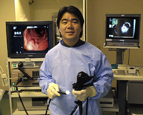
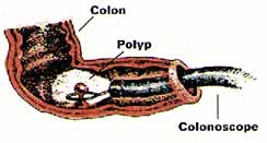
The instrument used to perform this test is the colonoscope: a long, flexible tube about the width of your index finger. Within the end of this tube is a miniaturized color-TV camera with a wide-angle lens. After passing this "scope" through the rectum and into your colon, your doctor then directly examines the lining of your lower digestive tract on a television monitor. In this manner, it is possible to evaluate intestinal inflammation, ulceration, bleeding, diverticulitis, colitis, colon polyps, tumors, etc. As part of your assessment, you may have undergone a barium enema examination of the colon, or "lower GI series." This technique, which utilizes x-rays, is helpful in identifying any areas of possible abnormality. X-rays are only shadows, however, and often do not fully demonstrate what is wrong. For example, the barium enema x-ray may miss nearly forty percent of colon polyps.

In the past, you also may have undergone a proctoscopic examination of the lower colon and rectum. Performed with a short, rigid, hollow tube, this limited procedure enabled the doctor to examine only the last ten inches of the large intestine. In most offices, the proctoscope has now been replaced by the flexible sigmoidoscope. This newer device enables your doctor to examine the last two feet of the colon while its flexibility affords the patient much greater comfort than does the rigid type of scope. The colonoscope offers a still greater advantage. With this longer flexible instrument, your doctor is usually able to directly examine the entire length of your colon - all five feet of its numerous twists and turns. In this manner, your doctor can be most certain about the condition of your colon.
..


How You Can Help
Your cooperation is essential for a successful examination. There are certain things you can do to help before the test begins:
  • Preparation A colonoscopy exam requires a thorough cleansing of the large intestine so that nothing impedes your doctor's view. The colon must be completely free of all solid waste. Since particles of stool can interfere with the examination, you will require a special dietary and laxative preparation on the day prior to the test. To be sure that you fully understand all aspects of the preparation, the GI Assistant or a member of the Nursing staff will review the preparation with you. It is critical that you follow the instructions as given. If the colon is not clean, the test - as well as the preparation - may have to be rescheduled. A few hints: It is best not to be constipated before the laxative preparation. This may lead to a temporary backup of the laxative solution, cramps, bloating, nausea, and vomiting. If you tend to be constipated, take one or two ounces of Milk of Magnesia the day before the laxative prep to "open up your pipes." This will make the actual preparation easier. If you develop mild distress during your prep, simply stop it for an hour or two and then resume it where you left off. If you develop severe abdominal cramps or persistent nausea and vomiting, stop the prep and call our office to reach our doctor on call. Of course, the laxative preparation will cause you to have many bowel movements. This may cause some rectal irritation and a small amount of bleeding. Many patients find that using Charmin Plus toilet tissue minimizes such rectal irritation. A nonprescription 1% hydrocortisone cream may also be soothing when applied to the rectal area after each bowel movement. Please avoid foods with many small seeds for a few days before the exam. They tend to linger in the colon and can clog our instruments. In addition, you must avoid all red-colored Jello products during your preparation as their red dye masks the lining of the colon.
  • Medications Medications containing iron or Metamucil-like fiber can impair your doctor's view of the colon. If possible, they should be temporarily discontinued several days before the test. If you are taking chronic aspirin or Persantine therapy, diabetic medication, or the blood-thinner Coumadin, you will require special preparation and adjustment of your medicines before the test. Please call our office for instructions. All other routine medications may continue to be taken with a small sip of water, even on the day of the exam.
  • What To Wear You will have to change into a patient gown before the examination. If this test is being done on an outpatient basis, you should wear, loose, comfortable, casual clothing that is easily removed and folded. Avoid girdles, pantyhose, or tight-fitting garments. Please leave your jewlery, valuables, and high heels at home. Transportation To minimize any discomfort, you will receive an injection before the colonoscopy. As this medication will make you drowsy for several hours, you cannot safely drive a car for the remainder of the day. Therefore, if this test is being done on an outpatient basis, a family member or friend must accompany you to the office in order to drive you home. If possible, you should choose someone with whom the doctor can freely discuss the results of your test. We request that your companion remain in the office during the test. You should arrive several minutes before your appointment and plan to stay approximately two hours. If the test is being done on an inpatient basis, a driver will not be needed. After the test, you will simply return to your hospital room to sleep off the remainder of the medication. You will be asked to remain in bed several hours to allow the sedation to wear off completely.
  • Your Permission If you have any questions or concerns about this test, do not hesitate to ask your doctor about them. To signify that you completely understand what this test involves, you will be asked to sign a written consent form, or "permit," before the test begins.

Looking Inside
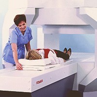
Prior to the test, you will be asked to remove your clothing and to slip into a patient gown. After signing the permission form, you will be positioned comfortably on your left side on the padded colonoscopy table. A small painless oximeter probe will be placed on your fingertip to monitor your pulse rate and breathing function during the exam. A thin nasal oxygen tube may be used to administer low flow oxygen during the exam. Then you will be given an intravenous injection of Demerol (pain killer) and Versed (tranquilizer); these medications will make you drowsy and relaxed, thus minimizing any discomfort during the examination. Please inform the doctor if you are allergic to Demerol or Versed, or if you chronically take narcotic painkillers such as Percodan, Percocet or Codeine, so that other medications can be substituted. Furthermore, rest assured that these medications will not prompt you to act or speak foolishly during the test.
..
Your doctor will then examine and lubricate the rectal opening to allow simple passage of the colonoscope. This will not be painful, but is mildly uncomfortable for a few seconds. After the colonoscope is properly positioned, your doctor gently guides the scope through the colon, a process that takes approximately one-half hour. To improve visualization, your doctor gently infuses air into the colon. This may cause a sensation of abdominal fullness, but should not be painful. In fact, many patients actually fall asleep during the examination. As the test progresses, you may be asked to change your position now and then to facilitate the passage of the colonoscope through the colon.
Here is an xray of the abdomen taken during a colonscopy which demonstrates how far the scope travels into the colon all the way to the right side of the colon.
..
Occasionally, passage of the colonoscope through the entire colon is impossible. While this may be caused by a blockage, it is most often caused by an extra "loop" of colon, adhesions from previous abdominal surgery, or severe diverticulosis. In such cases, a limited examination may be sufficient if the area of suspected abnormality has been visualized. If not, a barium enema x-ray (lower GI) may also be necessary.
..
Taking Specimens
If colonoscopy reveals any unusual condition such as inflammation, an ulcer, a tumor, or a polyp, your doctor may photograph it. This photo provides a permanent record for your medical chart and allows your other doctors to see what is wrong. With the advent of video-colonoscopy, it is now also possible to record the examination on a videocassette for later review when necessary. To better evaluate any areas of suspected abnormality, your doctor may take a brushing or biopsy of the colon lining. A brushing involves the passage of a tiny nylon brush through the center of the colonoscope. The brush rubs against the lining of the colon and retrieves bits of tissue for later analysis. To take a biopsy, the doctor passes a metal forceps through the scope and snips off a tiny sample of colon tissue for laboratory analysis. Both procedures are totally painless for the patient. Be assured that the decision to take a biopsy does not necessarily mean that your doctor suspects cancer. Biopsies of the digestive tract are often taken to look for other problems such as inflammation and ulcers.
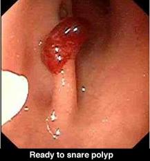
..
Removing Colon Polyps
The lining of the colon and rectum is normally smooth. Sometimes, however, growths known as "colon polyps" appear on the lining of the large intestine. Varying in size from pinheads to growths several inches in diameter, polyps do not usually produce any symptoms. Contrary to popular belief, polyps affect both men and women equally. Most colon polyps are initially non-cancerous. Nevertheless, as colon polyps continue to grow, they often develop into cancer of the colon. Colon cancer has become quite common in our society. In fact, nearly 1 in every 20 adult Americans will develop colon cancer in their lifetime. Colon cancer is now the leading cause of cancer deaths in nonsmokers. Only lung cancer takes a greater toll. It is not widely known that colon cancer now kills more women than breast cancer and more men than prostate cancer. But, there is good news. Research has confirmed that the single best prevention for colon cancer is the early detection and removal of all colon polyps!!! In the past, colon polyps located beyond the range of the proctoscope could only be removed by a major abdominal operation. Of course, this procedure required about a week of hospitalization and over a month of time lost from work for recovery. Fortunately, medical technology now permits the simple removal of most colon polyps. Should a small polyp be discovered during your colonoscopy, the polyp can usually be removed immediately. This prevents a potential colon cancer while eliminating the need for major surgery.
..
The doctor removes the polyp quite simply by placing a wire loop, or "snare," around the polyp's base and slowly tightening the loop. As the polyp is severed, a mild electric current cauterizes the tissue to prevent bleeding. There is no pain or any sensation as the polyp is removed. Polyp removal can usually be done on an outpatient basis. Moreover, since there is no incision, you may usually resume normal activity the next day.
..
After The Test
After an outpatient colonoscopy, you will be asked to rest awhile in the recovery room. Your companion will be asked to sit with you while the effects of the sedative begin to wear off. Once you are more alert, the doctor will meet with you to discuss the findings and any needed treatment. You will then be able to return home with your companion's assistance. However, since you will still be somewhat drowsy and uncoordinated, you will be taken directly to your car in a wheelchair by the office personnel. It is important that you go directly home; do not stop to eat along the way. Although you may resume a regular diet at home, you should eat lightly at first, and then gradually increase your intake of foods as tolerated. Since air was placed in the colon during the examination, you may experience the discomfort of mild "gas pains" for several hours until the gas is expelled. Because of the sedation's lingering effects, you should not drive, operate any machinery, drink alcohol, or engage in any vigorous activity for the remainder of the day. If the test is performed on an inpatient basis, you will be taken back to your hospital bed for recovery. If any specimens or polyps were extracted during the examination, the doctor will contact both you and your personal physician when the laboratory results become available. Further treatment or tests, if necessary, will be discussed at that time.
..
Is This Test Dangerous?
While every medical procedure involves some degree of risk, complications rarely occur in patients undergoing colonoscopy. With the advent of flexible fiberoptic instruments, this test has become a safe and simple method of directly examining the lower digestive tract. When performed by a physician who is specially trained and experienced in the procedure, the benefits of colonoscopy far exceed the risks.
..
Your doctor is a Gastroenterologist. In addition to standard medical training, he has received special instruction in diseases of the digestive system and has been thoroughly trained in the safe and proper operation of the colonoscope. The combination of his expertise and your cooperation should make this test as safe and simple as possible. However, as with all medical procedures, complications can occur.
..
The principal risks are perforation of the colon (a tear through the bowel wall) or bleeding. Although perforation generally requires surgery, certain cases may be treated with antibiotics and intravenous fluids. Bleeding may occur at the site of either a biopsy or polyp removal. Typically minor in degree, such bleeding may stop on its own or be controlled by cauterization. Occasionally, surgery is necessary. Fortunately, both perforation of the colon and bleeding are quite rare. Because bleeding may sometimes occur up to three weeks after a colon polyp has been removed, you should not plan to travel to any remote areas without medical access during this period of healing.
..
Other possible risks include drug reactions and complications related to other diseases that you may have. Consequently, you should inform your doctor of all allergic tendencies and medical problems. Occasionally, the site of the sedative injection may become inflamed and tender for a short time. This is not serious. If inflammation occurs, applying warm compresses to the area is usually helpful. While any of these complications may occur, it is well to remember that each of them occurs quite infrequently. Your doctor will be happy to discuss the above risks with you further, especially with regard to your particular situation and need for colonoscopy.
..
How About AIDS?
Due to the recent extensive coverage of AIDS by the media, some patients have expressed the concern that they might, in fact, contract AIDS through this examination. Be assured that this is not the case. All our instruments are thoroughly cleaned and undergo high-level disinfection after each use. Only techniques known to kill all disease-causing bacteria and viruses, including the hepatitis and AIDS virus, are employed in this process.
..
A Valuable Tool....
In summary, colonoscopy is a valuable tool for the diagnosis and treatment of many diseases of the large intestine. Even when x-rays are normal, the cause of symptoms such as rectal bleeding or change in bowel habits may be determined by colonoscopy. It is also useful in the diagnosis and follow-up care of patients with colitis. Through colonoscopy, the detection and removal of colon polyps, and thus the prevention of colon cancer, are made possible. Periodic colonoscopy is a valuable tool in monitoring patients with previous polyps, colon cancer, or a family history of colon cancer. In essence, colonoscopy is a safe and worthwhile procedure that is extremely well tolerated. If you have any questions about your need for colonoscopy, do not hesitate to ask your doctor.


To view information on another disease, click on SOD and Pancreatitis Library.!

SOD and Pancreatitis Library


..

Flexible Sigmoidoscopy

Flexible Sigmoidoscopy
..
Your doctor may have recommended that you undergo a special examination of your colon called Flexible Sigmoidoscopy, commonly called a "flex sig" Since your cooperation is essential for a successful and complete examination, it is important that you know exactly what is to be done and why. If, after reading this information, you have any additional questions concerning this procedure, ask your doctor.
..
What Is Flexible Sigmoidoscopy?
In the past, you may have experienced or heard of a proctoscope examination of the lower colon and rectum. This was performed with a short rigid hollow tube which allowed your doctor to examine only the last 10 inches of the large intestine. This device was often painful and, unfortunately, gave colon checkups a bad reputation as a very uncomfortable and embarrassing examination. Fortunately, recent progress in the field of fiberoptics has made colon exams much easier and more accurate. A new device, called a flexible sigmoidoscope, a 24 inch long thin flexible tube which contains a tiny color video camera is now used. This allows your doctor to perform a much more accurate and comfortable examination than the old "procto."
..
Why Do I Need This Test?
Flexible sigmoidoscopy can be done for a number of reasons, but the most common one is to help prevent colon cancer. It is now known that most colon cancers begin many years before as a small noncancerous growth called a polyp. But unfortunately, polyps cause no symptoms until they develop into colon cancer and by then it is too late. To minimize your personal risk of colon cancer, your colon must be periodically examined - just as one has periodic mammograms and prostate checkups. If a polyp is found during this exam and later removed, a potential colon cancer can be prevented. In other circumstances, flexible sigmoidoscopy can also be done to examine the left colon for signs of colitis, or to further evaluate symptoms such as diarrhea and rectal bleeding.
..
Is This Test Dangerous?
There is some risk to any medical procedure, but the frequency of serious complications during flexible sigmoidoscopy is quite low. When performed by an experienced physician, the benefits far exceed the risks. The combination of your doctor's experience and your cooperation should make this test as safe and simple as possible. However, as with all medical procedures, complications can occur. The principal risks are perforation of the colon (a tear through the bowel wall) or bleeding. A perforation usually requires surgery but may be managed with antibiotics and intravenous fluids in selected cases. Bleeding from flexible sigmoidoscopy is usually minor and stops on its own or can be controlled with treatment. Occasionally, blood transfusions and surgery are necessary. Fortunately, both of these problems are quite rare during flexible sigmoidoscopy - about 1 case in 8000 for perforation and 1 case in 17,000 for bleeding.
..
Scope Disinfection
With all the attention that AIDS and Hepatitis C have had in the media, some individuals have been concerned that they might contract a disease from these examinations. Be assured that this is not the case. All of the instruments are extensively cleaned and completely disinfected between each case with techniques that have been shown to effectively kill disease-causing bacteria and viruses including those which cause AIDS and hepatitis.
..
How Do I Prepare For The Exam?
Your cooperation is essential for a successful and complete examination. There are certain things you can do to help before the test begins. Your colon must be empty during flexible sigmoidoscopy so that your doctor's view is not blocked by particles of stool. This will require a special dietary and laxative preparation the day before the test. Detailed preparation instructions will be provided by the office staff. Be sure your doctor knows if you are taking insulin injections or Coumadin anticoagulants. Unless otherwise instructed, you may take all of your other routine medications as usual, even the day of the exam. Since anesthesia is not needed, a driver is not required.
..
Looking Inside
On the day of examination, wear comfortable easily-folded clothing that is simple to remove. Prior to the test, you will be asked to remove your clothing and to slip into a patient gown. Then, after a consent form is signed, you will be positioned comfortably on your left side on the padded examination table. The doctor will then do a rectal exam with a gloved, lubricated finger; then the lubricated scope will be gently inserted. This will not be painful, but is mildly uncomfortable for a few seconds. After the scope is positioned, the doctor will gently guide the flexible scope into the colon and examine the lining. To improve his view, your doctor will gently infuse air into the colon. This may cause a temporary sensation of abdominal fullness, cramping, or "gas," but the entire procedure should take less than three minutes. After The Test After the exam, you will be allowed to dress and have a glass of juice. The doctor will then explain to you exactly what was found and if any further tests or treatment will be necessary. Since this test does not require anesthesia, you will be able to immediately resume a regular diet and activities. Eat lightly at first, and then advance your diet as tolerated. Since air was placed in the colon during the examination, you may feel a little bloated and crampy until the extra air is expelled.
..
What If The Test Is Abnormal?
Don't panic. If a polyp is found, it is unlikely to be cancer - most are not. As a plus, most colon polyps can now be removed without the need for major abdominal surgery through a simple outpatient surgical procedure called colonoscopy. This more extensive "scope" examination is usually painless and does not require hospitalization. Most patients are back to work in a day or two. If more serious problems are found, the doctor will explain what medical or surgical forms of treatment are available.
..
What If The Test Is Normal?
Of course, since the flexible sigmoidoscope is only 2 feet long and the average colon is 5 feet long, not all of the colon is examined during this procedure. But, flexible sigmoidoscopy does allow examination of the area where the majority of colon cancer, polyps, and other problems are found. You must realize that this test is a simple screening exam and it is possible that problems further inside the colon may be missed. However, if you are in a low risk group, have no family history of colon cancer, no symptoms, nor signs of blood in the stool, the latest American Cancer Society guidelines merely recommend another flexible sigmoidoscopy every five years in the future and a stool test for hidden blood, or Hemoccult slide test, annually. However if you have symptoms or are in a higher risk group, your doctor may wish to alter these guidelines to your particular case. In Summary So as you have learned, flexible sigmoidoscopy is a safe, simple, and highly effective diagnostic technique that allows your doctor to directly inspect the lining of your left colon. As an individual, you can dramatically reduce your risk of getting colon cancer by having regular examinations before symptoms develop. If you are over 50 years old, consult with your family physician about periodic colon examinations. Prevention means taking charge of your health. Following these few simple steps can keep you healthy to enjoy the good life you have worked so hard to create. It's worth the effort....
..


To view information on another disease, click on SOD and Pancreatitis Library.!

SOD and Pancreatitis Library


..

MRI and MRCP

Magnetic Resonance Imaging (MRI)
...
What is an MRI?
Most radiology imaging studies use x-rays to visualize what lies inside the body. For example, a chest x-ray allows the doctor to see through the skin and study the heart and lungs. Modern x-rays are very safe, but do expose the body to some x-ray radiation. Magnetic resonance imaging (MRI) is a different method of looking inside the body. Instead of x-rays, the MRI scanner uses magnetism and radio waves to produce remarkably clear pictures. The powerful magnetic field causes the hydrogen ions in the body to become magnetized and line up in a certain order. The data received is analyzed and turned into an image by a high powered computer to create detailed image slices (cross sections) of your body. MRI can produce better soft-tissue images than standard x-rays and is better at distinguishing normal, healthy soft tissue from diseased tissue.
...
How does MRI differ from a CT scan?
One of the most basic differences between the two tests is that Computerized Tomography (CT) Scanning uses x-rays and MRI does not. A CT scan uses faster scanning times and can be performed in patients with pacemakers and other metallic implants. But a CT scan does expose the patient to x-rays and risks allergic reactions to intravenously administered iodine-containing dye. The MRI produces better images of the body's soft tissues and involves no x-rays or iodine dyes. But MRI scanning times are longer and difficult for patients who are not able to hold their breath. Patients with pacemakers and intra-cerebral aneurysm clips cannot be scanned by MRI.
...
Where Is an MRI performed?
MRI studies can be safely and accurately performed in a hospital radiology department, in a mobile MRI unit, or a freestanding MRI center.
..
Who performs the exam?
An MRI study is performed by a trained MRI technician under the supervision of a radiologist, a medical doctor trained in special imaging studies. The results of the test are analyzed by the radiologist and reported to your doctor within a few days.
..
Regular vs "Open" MRI
There are two types of MRI magnet machines: Closed and Open, based upon their shape. The standard "Closed" MRI is done in a narrow tube-like device about 2 feet in diameter and 6 feet to 8 feet long to optimize the images. Because of the small bore of the magnet, some patients experience claustrophobia and have difficulty in cooperating during the study. In the "Open" type, the large magnet that generates the image is generally suspended a couple of feet above the patient, and except for its supports, the unit is Open all around. If a patient is severely claustrophobic or over 300 pounds in weight, the doctor may suggest that the examination be done in an "Open" MRI unit because it has more room inside than a Closed magnet. Most Open units can accommodate patients up to 450 pounds in weight. The Open unit is more "patient friendly", but most radiologist feel that the Closed MRI operates much faster and produces a higher-resolution image with finer detail than the Open type. Closed magnets can be used for all MRI procedures, and Open magnets are used for more routine applications. Open MRI technology has improved over the years and may be adequate in many cases. Specialized studies such as MR angiography (MRA) and MR cholangiopancreatography (MRCP) can only be performed in Closed MRI machines.

Scheduling an MRI
An MRI is usually scheduled through the local hospital radiology department or a freestanding Open MRI center. In order to perform the study, they need an order for the study and an insurance referral from your physician. MRI is a non-invasive test and really without significant risks. One concern, however, is the "projectile effect", which involves the forceful attraction of metallic objects to the magnet. Because of this, there are several conditions under which MRI may not be safe. Patients should notify the receptionist or technologist prior to their appointment if they have any of the following:


  1. An implanted pacemaker, defibrillator ("AICD"), or heart valve
  2. An implanted pump device (such as an insulin or pain medication pump)
  3. An inner ear implant
  4. An aneurysm clip within the brain
  5. An intrauterine device (IUD)
  6. Metal in the eyes (at any time), or have ever been a metal worker of any kind
  7. Permanent tattoo eyeliner
  8. Currently pregnant
  9. Artificial joints or metallic plates
  10. Shrapnel Patients can safely undergo MRI with orthopedic hardware in their joints, such as a metallic plate or hip replacement. However, if the metal device is located close to the part of the body being examined, the images can be seriously degraded and useless.
..
MR cholangiopancreatography (MRCP)
This big word refers to a special test of the liver, bile ducts, and pancreas done using MRI. MRCP can produce images very similar as those obtained from the more invasive approach with ERCP (Endoscopic Retrograde Cholanigiopancreatography) without the added risk of pancreatitis, sedation, and perforation. However, image quality is less with MRCP and there is no way to correct whatever problem is found, as there is during ERCP. ERCP is best used when there is a high likelihood of gallstones obstructing the bile ducts or another blockage of the liver or pancreas. MRCP is of value in patients with a low probability of gallstones or obstruction in the bile ducts or pancreas, or in patients who are too sick for the anesthesia required for ERCP.
..
Preparing for an MRI
There is no special preparation for an MRI examination. There is no need for a change in daily routine. All prescription medications can be taken normally. However, patients undergoing MRI examination of the gallbladder and bile ducts (MRCP) will be asked to not eat for 12 hours prior to imaging. No special preparation is required for other body examinations. Patients are asked to bring the physician's order, insurance cards, referral forms, and any previous MRI, CT, or x-ray films relating to their exam. It is best to wear loose clothing without zippers or metallic parts. Elastic waistbands are suggested. During the exam The MRI technician explains the exam and answers any questions the patient may have. The patient may be asked to sign a consent form giving permission for the test and may be asked to change into a patient gown. Because MRI uses a powerful magnet, watches, metal objects in pockets, and credit cards with magnetic strips will not be permitted in the MRI room. Patients must also remove any other metallic objects such as jewelry, hairpins, eye glasses, wigs (if it has metallic clips), and non-permanent dentures. In a Closed MRI unit, the patient is positioned on a scanning table, head first, with arms at the side. The scanning table then slides into the magnet, covering the whole body. For clear pictures, the patient will be asked to hold very still, and in some cases, to hold their breath for up to 30 seconds. There is no pain or other sensation during the exam; however, an MRI is a noisy machine which produces intermittent humming, clicking and knocking sounds. Earplugs are available. Most MRI units also provide an assortment of music to help the patient relax. Patient are welcome to bring their own CD or cassette. There is a two-way intercom providing communication between patient and the technologist. For some studies such as MRCP, the radiologist will inject a "contrast agent" into a vein to improve the quality of the images. This material is injected into a vein in the arm. How long does an MRI exam take? The length of MRI examinations can vary from 15 minutes to 1 1/2 hours, averaging 45 minutes. Each test consists of several sequences or collections of data gathered over 2 to 10 minutes. After the exam There are no post-exam instructions. You may resume your normal diet and activities.
..
What about pregancy?
Although there are no known side effects of magnetism on the developing baby, it is recommended that a pregnant woman wait until the second trimester for MR imaging.

Getting the test results?
Because very large amounts of data are created during these studies, they can easily have hundreds of images that require hours of manipulation to interpret. The study will be read by a board-certified radiologist who sends the results to the referring physician who will notify the patient. Results are usually available within 72 hours.
..
What does an MRI cost?
The cost of an MRI study can range from $400 to more than $2,000, with a typical cost being about $800. Most health insurance, including Medicare, covers MRI testing. In summary MRI stands for Magnetic Resonance Imaging. An MRI offers a safe and efficient method of diagnosing many conditions, without the use of harmful x-rays. In many cases, MRI can lead to early detection and treatment of disease without surgery or biopsy. It is a non-invasive method of examining the soft tissue of the body including organs, muscles and tendons and requires little patient preparation. If you have any more questions about MRI, ask your doctor.
..
..

Magnetic Resonance Cholangiography (MRCP)
By: A. Alan White, M.D.
..
MRCP is a safe, non-invasive, easily performed and reliable procedure for evaluation of the bile ducts and the main pancreatic duct. No injections are required since the MR technique images the fluid normally present in the ducts. This is a procedure commonly utilized in major teaching institutions around the world.
..
Good quality examinations require a high field magnet, strong, fast gradients, special coils, and software that allows obtaining breath-hold heavily T-2 weighted images. All fluid containing structures will appear bright on these exams.
..
Techniques:
The patient fasts for 4 hours prior to the study to allow filling of the gallbladder and emptying of the stomach. The patient usually drinks about 100cc of a non absorbable iron containing liquid to decrease the signal of fluid in the stomach. The patient does not need IV contrast agents and there is no radiation exposure.
..
A series of images is then obtained during breath-holding periods of 2 to 25 seconds. Since the images are obtained rapidly, a series of images taken every few seconds through the distal duct can demonstrate contraction and relaxation of the distal common duct and sphincter of Oddi.
..
MRCP is based on a heavily T2 weighted pulse sequence which shows stationary fluids, such as bile, to tappear at high signal intensity whereas the surrounding liver and flowing blood generates little signal. As a result of this combination of imaging characteristics, MRCP provides optimal contrast between the hyperintense signal of the bile and the hypointense signal of background tissue.
..
Accuracy:
Detection of stones in the common duct by MRCP has an accuracy in the 90-95% range which is very comparable to ERCP. The accuracy of detection of the point of benign or malignant obstruction of the biliary duct or ducts is in the 90% range. Diagnosis of chronic pancreatitis involving the main duct is very comparable to ERCP with 90% correlation.
..
Indications for MRCP:

  • Patients where ERCP has failed, where ERCP carries a high risk and in patients where prior surgery makes access difficult are best studied by MRCP. In patients with low to moderate clinical suspicion of bile duct stones MRCP should be considered because of its high accuracy and lack of risk. With a high clinical suspicion of bile duct stones, ERCP is a logical choice since therapy can be instituted at the time of the procedure. Patients with acute pancreatitis have an increased risk of complication from ERCP and in patients with a low to moderate clinical suspicion of bile duct stones MRCP offers a no risk alternative.
  • Demonstration of ductal anatomy above a point of complete obstruction is another use of MRCP and is helpful in planning therapeutic intervention for bypass and drainage.
  • Suspected chronic pancreatitis, demonstration of anatomic variants such as pancreas divisum, aberrant cystic duct and other duct anomalies, sclerosing cholangitis, choledochal cysts, anastomic strictures and demonstration of post surgical anatomy are all uses for MRCP. Pre-operative evaluation of patients prior to laparoscopic cholecystectomy is currently under study. MRCP would be useful in those 10-15% of patients with common duct stones in helping plan the surgical procedure.
..
Advantages of MRCP Compared to ERCP:

  1. Non invasive (avoids complications of diagnostic ERCP or PTC)
  2. Usually no sedation required
  3. No iodinated intravenous contrast
  4. Rapid scan time
  5. No ionizing radiation
  6. Delineates ductal anatomy proximal to obstruction
  7. Delineates anatomy post biliary-enteric anastomosis
..
Contraindication:
The only contraindications are those of MRI in general including cardiac pacemakers, ferromagnetic aneurysm clips, intraorbital metallic foreign bodies and severe claustrophobia.
..
Summary:
MRCP has high diagnostic accuracy, is completely safe and avoids the risk of pancreatitis, bleeding and perforation associated with invasive techniques. The exam is well tolerated by most patients and diagnostic quality images can be obtained 99% of the time.
..


To view information on another disease, click on SOD and Pancreatitis Library.!

SOD and Pancreatitis Library


..

Barium X-rays, Ultrasound (Sonogram), Cat Scans, Nuclear Scans

Special Imaging Studies
...

Your doctor may have recently requested that you have an examination of your digestive tract at your hospital outpatient radiology (x-ray) department. If you are scheduled to have such an exam, it is important that you understand why the test is necessary and what steps are needed for proper preparation. This information reviews the most common studies performed. If you have any additional questions, please ask.
...
Why Do I Need This Test?
Tests such as these are done to provide your doctor with additional information about the status of your digestive system. This helps your doctor decide what, if any, problems may be present, and the best remedy in your particular case.
  1. Often, x-rays are requested to evaluate an organ that cannot be seen by other means. For instance, a diseased gallbladder may not show up in blood tests, nor can a standard "scope" test see this organ. But, specialized x-rays and other studies can visualize the gallbladder and determine if stones or disease are present.
  2. Sometimes x-rays are used to visualize part of the intestine that cannot be reached with a scope exam. A common example would be a barium x-ray of the portion of the small intestine that lies beyond the reach of a upper or lower scope test.
  3. Occasionally, a barium x-ray of the colon is ordered when severe diverticulosis or other problems prevent completion of a full colonoscopy "scope" examination.
..
If you need to have x-rays or other special studies performed, your doctor will explain what tests are necessary and why.
..
Here is a summary of the four basic exams:
  1. Barium X-rays You may not realize that when ordinary x-rays are taken, only your bones show up. X-ray beams pass right through the soft tissues of your body, such as the intestines, liver, kidneys, etc., making them almost invisible. Barium is an inert, harmless mineral that prevents the passage of x-rays - just as your hand blocks sunlight. During an Upper Gastrointestinal Series, or Upper GI Series, you are given a flavored "milkshake" of liquid barium to drink. The barium fills your stomach making it visible on the x-ray film. The standard Upper GI evaluates the esophagus, stomach, and first portion of your small intestine - called the duodenum. This takes about 30 minutes. If your doctor requests that the remaining twenty feet of small intestine also be evaluated, the test is termed a Small Bowel Series and takes an additional two to three hours to complete. Both examinations require fasting after midnight, but no laxative preparation is necessary, nor is a driver needed. A similar examination of the large intestine, or colon, is called a Lower Gastrointestinal Series, or Lower GI. Of course, in this instance, the barium is not swallowed, but is given rectally as an enema - thus the more common name, Barium Enema. This examination takes less than an hour and requires fasting as well as an unpleasant laxative and dietary preparation the day or two before. Prep instructions vary and will be provided by our staff. Be sure to follow them exactly so the test need not be repeated.
  2. Ultrasound Another test often performed in the x-ray department does not use x-rays at all - an Abdominal Ultrasound. Also known as a Sonogram, this test utilizes specialized sound waves that bounce off your internal organs to obtain images - much like the way a submarine uses sonar to locate the enemy. The pitch, or frequency, of these sound waves is far above the range of human hearing, hence the name Ultrasound. An Ultrasound is also the examination that is given to pregnant women to check the size of their baby before delivery. This same exam can also be used to visualize certain digestive organs, such as the gallbladder, liver and pancreas as well as the main abdominal blood vessel, the aorta. During an ultrasound, you will be positioned on an examination table and a gel will be applied to your abdomen. A small probe, called a transducer, will be passed over the surface of your abdomen. The test is quick, painless, and requires very little preparation. Fasting after midnight is usually required. A driver is not necessary. If your doctor also requests an evaluation of the lower abdomen, or pelvis, a full urinary bladder is also required.
  3. CAT Scans A CAT Scan, also known as CT Scan, uses a complex computerized x-ray scanner that takes multiple views of your abdominal organs as you lie on a flat table. A high speed computer analyses the information to create cross sectional images of parts of your body. The radiologist (x-ray doctor) can then view these images in sequence, like slices in a loaf of bread, and create a virtual three dimensional view of your abdominal organs. CT is best suited for viewing the more solid digestive organs like liver, pancreas and kidney, and less suited for viewing the hollow air field intestinal tract. Preparation is simple, however there are a few concerns. First, since dehydration can increase the risk of side effects, it is important that you drink plenty of fluids up until four hours before the examination. During the last four hours, you should avoid all food and drink. Since the contrast material can worsen previously existing kidney disease, the radiologist usually requests proof of normal kidney function. This requires a blood test. To make the examination more accurate, you will be asked to drink an oral contrast solution before the scan. Additional contrast material may also be given by vein to enhance the images. Since this solution contains iodine, be sure to tell the doctor if you have ever had an allergic reaction to iodine, IVP or catheterization dye, or shellfish. Depending on the nature of your previous reaction, the solution will have to be modified or simply not used. The CT scan itself is simple and painless, and usually takes less than an hour to complete. Serious side effects are rare, but a temporary feeling of warmth and mild nausea are common after the contrast injection. After the examination, you will be able to resume your normal activities and diet. You should drink plenty of fluids for 24 hours after the examination to help flush out your system.
  4. Nuclear Scans In some instances, your doctor may request that you have a nuclear scan. Although the name sounds a little unsettling, a nuclear scan involves only a small "tracer" dose of radioactive material, and is not dangerous. Once this tracer is injected into your system, it can be followed through your digestive organs as you lie directly underneath a large, flat Geiger counter. A nuclear scan is most often used to assess liver and gallbladder function. Other uses include measurement of stomach emptying and localization of intestinal bleeding. Nuclear scans require very little preparation.

..

Where Is The Test Performed?
X-ray, Ultrasound, CAT scan and Nuclear Scan exams require specialized equipment and highly trained technicians. They are usually performed in a hospital our specialized outpatient facility.
..
Who Performs The Exam?
In most cases, the examination will be performed by a specially trained technician who is expert in using the equipment. The radiologist may or may not be present depending on the circumstances. However, the radiologist will review the final films and proceed with an official evaluation.
..
How Do I Schedule My Test?
Your doctor's office can assist you in obtaining all the necessary information to schedule your examination with your local outpatient radiology department. Any special preparation instructions will be provided.
..
Preparing for Your
Test Careful preparation is an important part of any examination. Preparation instructions vary from test to test and will be provided by our office staff. In all instances, you should wear easily-removable and comfortable clothing. Leave girdles, valuables, high heels, and jewelry at home. Be sure that you fully understand what preparation is required of you so that a safe and accurate examination can be performed. If you do not, the examination may have to be repeated at a later date. If you have any questions, please ask.
..
After Your Exam
After your examination is completed, you will be able to immediately resume normal activities and diet. You should drink plenty of extra fluids for the first 24 hours to help flush your system.
..
A word to the wise: If barium was used for your test, you might also wish to take a mild laxative such as an ounce of Milk of Magnesia after the x-ray. This helps eliminate the barium from your system. If this is not done, the barium can harden, much like plaster of Paris, making elimination quite difficult.
..
What About Side Effects?
There are no known side effects to health from ultrasound. Nuclear scans, CAT scans, and barium x-rays do expose your body to small amounts of radiation. However, the dose is quite low and felt to be generally safe. The exceptions would be pregnancy and breast feeding. Any woman who is pregnant, or thinks she might be, or is breastfeeding, should let her doctor know before scheduling the examination. Also, if you believe that you are allergic to iodine dyes and are scheduled for a CT scan, notify your doctor. How Do I Get The Results? Once the test is completed, the films are developed and analyzed by the radiologist who then sends us a written report. Your doctor will contact you after reviewing the results in the context of your case. This may take about a week, but most reports arrive sooner. In Summary In addition to "scope" tests, imaging techniques such as barium x-rays, Ultrasound studies, CAT and Nuclear Scans are often requested to obtain information about the status of your digestive system. These tests are generally safe and well tolerated, but require proper preparation. You have an important role in making your examination a success. If you have any questions, again please ask.
..


To view information on another disease, click on SOD and Pancreatitis Library.!

SOD and Pancreatitis Library


..

The Camera in a Pill!


Wireless Capsule Endoscopy - "The Camera in a Pill"
Searching For the Leak.... What if you were a plumber and couldn't find the leak? That is the situation that sometimes frustrates doctors asked to evaluate a patient with signs of iron deficiency anemia due to blood loss. There are many types of anemia. Iron deficiency anemia is a common type and most often occurs in pregnant women or menstruating women due to the monthly loss of blood. But when significant iron deficiency anemia occurs in women after the age of menopause or in men, it is often a sign of the loss of blood from somewhere else - usually the digestive tract. This blood loss may be very gradual and not visible to the patient. But over time, the continued loss of blood can deplete the body of iron and anemia develops.
If a doctor suspects that anemia is due to intestinal bleeding, special tests are usually ordered to help the doctor "find the leak." This is a bigger task than most people realize since the average adult digestive tract is approximately 30 feet in length. The top 4 feet includes the esophagus (foodpipe) and stomach and first portion of the small intestine, called the duodenum. The bottom 6 feet makes up the colon and rectum. In between, lies the rest of the 20 feet of small intestine where the process of digestion actually occurs.
..
Scope Tests
In this situation, one of the first diagnostic studies ordered are special "scope" tests of the digestive tract. Gastroscopy is used to check the first 4 feet of the upper digestive tract and colonoscopy to evaluate the colon and rectum. As you can see, most of the 20 feet of small intestine lies beyond the reach of these two studies. Fortunately, most bleeding problems seem to occur in the area than can be "scoped" and the source of bleeding is usually found and treated. Common problems would include Hiatal hernia, gastritis, ulcers, polyps, and, sometimes, stomach or colon cancer.
..
The Small Intestine - Uncharted TerritoryWhat if a patient had severe iron deficiency anemia and scope tests of the stomach and colon are normal? It is not uncommon for doctors to evaluate a patient with unexplained anemia and neither gastroscopy nor colonoscopy scope examinations reveal the diagnosis. By a process of elimination, it then becomes likely that the source of bleeding lies somewhere in-between - below the reach of the gastroscope and above the reach of the colonoscope - in the 20 feet of small intestine. How then is this area examined?
Well, not very well. Gastroscopy and colonoscopy cannot reach this far. Contrary to popular belief, special imaging studies like CT scan or MRI are not useful in this circumstance. X-rays of the small intestine can be performed after drinking a chalky solution of barium. Called a small bowel series, this test has been available for many years, but has a limited accuracy. X-rays are still only shadow pictures and do not view the object itself like a camera.
..
Why not use a longer scope?
Since scope tests were first invented, doctors have wanted to be able to visualize the entire gut - all 30 feet. But, a direct view of the small intestine has remained elusive. Attempts have been made to develop longer endoscopic instruments. This technique called push enteroscopy has had only limited success. The longer instruments are difficult to control and manipulate and are hard to maintain. The accuracy of push enteroscopy is still limited since even in the best of hands the entire small intestine is not visualized.
..
The Given Video Capsule - Hope For the Future
In 1981, an Israeli physician, Dr. Gavriel Iddan, began development of a video camera that would fit inside a pill. Technology was not ready and the idea was put on hold. It took 20 years for technology to catch up with Dr. Iddan. In 2001, the FDA approved the Given Diagnostic Imaging System. This may sound like science fiction, but this 11 x 26 mm capsule weighs only 4 gms (about 1/7th of an ounce) and contains a color video camera and wireless radiofrequency transmitter, 4 LED lights, and enough battery power to take 50,000 color images during an 8-hour journey through the digestive tract. About the size of a large vitamin, the capsule is made of specially sealed biocompatible material that is resistant to stomach acid and powerful digestive enzymes. Another name for this new technique is Wireless Capsule Endoscopy.
..
Patients report that the video capsule is easier to swallow than an aspirin. It seems that the most important factor in ease of swallowing is the lack of friction. The capsule is very smooth, enabling it to slip down the throat with just a sip of water. After the Given M2A capsule is swallowed, it moves through the digestive track naturally with the aid of the peristaltic activity of the intestinal muscles. The patient comfortably continues with regular activities throughout the examination without feeling sensations resulting from the capsule's passage. During the 8 hour exam, the images are continuously transmitted to special antenna pads placed on the body and captured on a recording device about the size of a portable Walkman which is worn about the patient's waist. After the exam, the patient returns to the doctor's office and the recording device is removed. The stored images are transferred to a computer PC workstation where they are transformed into a digital movie which the doctor can later examine on the computer monitor. Patients are not required to retrieve and return the video capsule to the physician. It is disposable and expelled normally and effortlessly with the next bowel movement.
..
This technique is very new, but preliminary reports have already demonstrated cases in which the Given videocapsule was able to make a diagnosis not seen by conventional studies. This remarkable device will never be 100% accurate. No medical procedure is. But current estimates suggest an accuracy of about 55%, an improvement over push enteroscopy. As of May 2002, the software is still evolving and there are concerns over insurance coverage, but this remarkable device shows great promise. Currently, it is not available through our center or in Western Pennsyvania, but hopefully, it will be in the future. For more information about this device and its availability, access the Given Imaging Ltd. Website at:

www.givenimaging.com


To view information on another disease, click on SOD and Pancreatitis Library.!

SOD and Pancreatitis Library


..

Hemoccult Card Test

Hemoccult Card Test (for hidden blood in the stool)

Tina: How a visit to her gynecologist saved her life.... Tina F. is a 52 year old school psychologist who sees her gynecologist every year for a routine checkup. Like many women, she does not have a family doctor, but uses her gynecologist as her personal physician. This year her doctor suggested that a routine stool Hemoccult test be performed in addition to her annual blood tests and physical exam. As she left the office, the nurse gave her a Hemoccult card test kit and instructions. Tina collected the stool specimens as instructed and mailed the card back to her doctor's office for testing. She was surprised to receive a letter from her doctor the next week informing her that the Hemoccult test found traces of hidden blood in her stool.
Tina was especially surprised since she never had any problems with bowel movements and never noticed any blood in her stool. She was referred to our office and a colonoscopy exam was performed to determine the cause of the bleeding. The colonoscopy exam went well and found the cause of Tina's bleeding - an inflamed colon polyp that was located in her right colon. It was slowly oozing small amounts of blood and was the source of the trace of hidden blood in the stool. In this series of photos, you can see how the polyp was removed during colonoscopy using a thin wire electrocautery snare. It was a simple outpatient procedure and she was back to work the next morning. Tina was pleased that her problem could be cured with a simple painless outpatient procedure and she was back to work the next morning. The polyp was sent for a biopsy.
We called Tina the next week and informed her that she was very lucky. The biopsy showed that her polyp was "highly dysplastic with carcinoma-in-situ bordering on adenocarcinoma" - about as close as you can get to cancer, without actually being malignant. Her gynecologist did her a great service by being thorough and discovering her rectal bleeding even before it was visually apparent. The discovery and removal of this type of colon polyp saved Tina from having to deal with colon cancer in her future and probably saved her life. She will be scheduled to return for a follow-up colon exam periodically in the future.
...
What is a Hemoccult test? The Hemoccult test, a product of SmithKline Diagnostics, Inc., is a simple screening test which detects blood in the stool (bowel movements).
It is a very sensitive test and can detect a small trace of blood even before it is visible to the naked eye. In medicine, the prefix "heme" refers to blood, as in "hematology". Blood which is hidden from view is called "occult" bleeding (nothing to do with witchcraft), hence the name Hemoccult for "hidden blood".

Other tests for occult blood are also on the market (Colocare, EZ Detect), but the most commonly used test is the Hemoccult.
..

Prevention - the purpose of the stool Hemoccult test is to help prevent colon cancer by searching for hidden (occult) blood in the stool before it has become so apparent to be visible. The American Cancer Society estimates that over 130,000 people developed colorectal cancer in 2000. Not only is this a common disease, it is quite sneaky. In fact, the majority of patients with colon cancer are incurable when they have their first symptom. Despite undergoing the rigors of heroic treatment such as surgery, radiation, and chemotherapy, most paitients succumb to their disease.
..Why do this test?In advanced stages, the overall cure rate is quite low. Over 56,000 Americans died last year from colon cancer. However, when colorectal cancer is detected in an early localized stage, up to 85% of colorectal cancers can be treated successfully. When cancer is detected after symptoms appear and it has spread to other parts of the body, fewer than 7% may be treated successfully. This means that earlier detection could save thousands of lives each year. In some cases, the Hemoccult test will show hidden blood in the stool in early curable stages of colon cancer. Even better, as in this case, the Hemoccult test may be positive even before cancer cells have developed. How is it done? It is quite simple. To complete the test, you place a thin smear of a small stool specimen, taken from your toilet bowl, onto the window inside the test slides included in your kit. You do this twice from each of three consecutive bowel movements and mail the completed card back to your doctor's office in the special envelope.
..
To maximize the accuracy, some special precautions are suggested:


  1. Do not collect samples during, or until three days after your menstrual period, or while you have bleeding hemorrhoids or blood in your urine.
  2. Remove any cleaning or deodorizing agents from the toilet and flush twice.
  3. For 3 days before the test, avoid Vitamin C tablets, iron tablets, red meat (rare, cooked and processed beef, lamb and liver), raw broccoli, cauliflower, horseradish, parsnips, radishes, turnips and melons.
  4. Protect slides from heat, light and chemicals. Keep cover flap of slides closed when not in use.
  5. To be sure the the card and developing solution are fresh and active, a quality control strip is included on the back of each card. Normally, the developing solution turns this control strip blue which means the card can be trusted.

What happens after the Hemoccult card is returned?

  1. After the stool collection is complete, the Hemoccult card is mailed back to the doctor's office or lab in a special protective envelope for testing.
  2. Testing the card is quite quick and simple. The medical technician opens the flaps on the back of the card and applies a few drops of a special chemical developing solution onto each of the three stool specimens.
  3. POSITIVE TEST The developer turns the stool specimen blue - this is an abnormal result and means that blood is present in that stool specimen.
  4. NEGATIVE TEST If the stool specimen does not turn blue, then no hidden blood is present. This is a normal result.


Who should do a Hemoccult test?

All adults over 40 should do this test annually even if there are no symptoms. This really should just be part of an annual checkup from your family doctor or gynecologist.

Who should NOT do a Hemoccult test?

Anyone with visible rectal bleeding. This test looks for hidden blood in the stool - before it is visible to the naked eye. If an individual has visible blood in the stool, there is no reason to do a Hemoccult test. You already know that bleeding is present and needs to be evaluated.

What if it is abnormal (POSITIVE)? Don't panic! A positive Hemoccult test (one that shows blood) does not diagnosis cancer. It simply means that there is blood in the stool that is not supposed to be there. There are many possible causes for blood in the stool which have nothing to do with colon cancer such as hemorrhoids, fissures, ulcers, and, as in the above example, colon polyps. The important point is that when a Hemoccult test is positive for occult blood, you must see your doctor for further testing to determine the cause. The safest approach is to assume that the cause is colon cancer until proven otherwise. Most adults who have an abnormal stool Hemoccult test will be referred to a gastroenterologist for further evaluation. As in Tina's case, this will usually include an examination of the colon, or colonoscopy. If the colon test is normal, the doctor will sometimes suggest that the upper digestive tract be evaluated in a similar fashion by also performing a gastroscopy test. Usually, both "scope" exams can be done on the same visit.

What if it is normal (NEGATIVE)? That sounds reassuring, but perhaps falsely so. The Hemoccult test is a test for blood, not cancer. The surface of a colon cancer is delicate and may bleed easily. In fact, this is the very basis of the test itself. But, you should know that most colon cancers and polyps bleed intermittently. They may lose a small amount of blood one day and not bleed again for several days. Some don't bleed at all - especially in early curable stages. So, a normal (NEGATIVE) stool Hemoccult test misses about 60% of colon polyps and 40% of colon cancers. Not very reassuring. This is really a screening test that has some significance only if abnormal (POSITIVE). The Hemoccult test is meant to be just a part of colon cancer prevention, but must be supplemented with more ac curate examinations of the colon, such as a screening sigmoidoscopy, a screening colonoscopy, or a barium enema.

How accurate is the Hemoccult test?

Not very. As mentioned above, the Hemoccult test is very sensitive for finding occult blood in the stool when present. It does so about 98% of the time. But since colon cancer and polyps may not bleed during the time of stool collection, a normal result may miss colon cancer and polyps up to 60% of the time. However, doing a Hemoccut test is better than doing nothing at all - which is what most adults do. The Hemoccult will still pick up about 40% of cases that would have been missed if no testing was done. However, the Hemoccult test should not be relied upon as the sole screening method.

Summary

The Hemoccult test is a simple, inexpensive, non-invasive stool test that can be collected in the privacy of your home. It is not a test for colon cancer, but detects hidden (occult) blood in the stool before it becomes visible. A normal test should not be reassuring since it misses over half of the colon cancers and polyps. It is not meant to be used alone, but must be part of a comprehensive colon cancer screening program which include other screening exams. A positive test should not cause panic, but must be further investigated to determine the source of bleeding. Only diagnostic testing from a physician can determine the cause of the bleeding. Used correctly, the stool Hemoccult test can be an important aid in detecting cancer in its early stages of development. If you have further questions, discuss them with your doctor.


To view information on another disease, click on SOD and Pancreatitis Library.!

SOD and Pancreatitis Library


..