...
 What is an MRI?
Most radiology imaging studies use x-rays to visualize what lies inside the body. For example, a chest x-ray allows the doctor to see through the skin and study the heart and lungs. Modern x-rays are very safe, but do expose the body to some x-ray radiation. Magnetic resonance imaging (MRI) is a different method of looking inside the body. Instead of x-rays, the MRI scanner uses magnetism and radio waves to produce remarkably clear pictures. The powerful magnetic field causes the hydrogen ions in the body to become magnetized and line up in a certain order. The data received is analyzed and turned into an image by a high powered computer to create detailed image slices (cross sections) of your body. MRI can produce better soft-tissue images than standard x-rays and is better at distinguishing normal, healthy soft tissue from diseased tissue.
...
How does MRI differ from a CT scan?
One of the most basic differences between the two tests is that Computerized Tomography (CT) Scanning uses x-rays and MRI does not. A CT scan uses faster scanning times and can be performed in patients with pacemakers and other metallic implants. But a CT scan does expose the patient to x-rays and risks allergic reactions to intravenously administered iodine-containing dye. The MRI produces better images of the body's soft tissues and involves no x-rays or iodine dyes. But MRI scanning times are longer and difficult for patients who are not able to hold their breath. Patients with pacemakers and intra-cerebral aneurysm clips cannot be scanned by MRI.
...
Where Is an MRI performed?
MRI studies can be safely and accurately performed in a hospital radiology department, in a mobile MRI unit, or a freestanding MRI center.
..
Who performs the exam?
An MRI study is performed by a trained MRI technician under the supervision of a radiologist, a medical doctor trained in special imaging studies. The results of the test are analyzed by the radiologist and reported to your doctor within a few days.
..
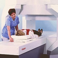
Regular vs "Open" MRI
There are two types of MRI magnet machines: Closed and Open, based upon their shape. The standard "Closed" MRI is done in a narrow tube-like device about 2 feet in diameter and 6 feet to 8 feet long to optimize the images. Because of the small bore of the magnet, some patients experience claustrophobia and have difficulty in cooperating during the study. In the "Open" type, the large magnet that generates the image is generally suspended a couple of feet above the patient, and except for its supports, the unit is Open all around. If a patient is severely claustrophobic or over 300 pounds in weight, the doctor may suggest that the examination be done in an "Open" MRI unit because it has more room inside than a Closed magnet. Most Open units can accommodate patients up to 450 pounds in weight. The Open unit is more "patient friendly", but most radiologist feel that the Closed MRI operates much faster and produces a higher-resolution image with finer detail than the Open type. Closed magnets can be used for all MRI procedures, and Open magnets are used for more routine applications. Open MRI technology has improved over the years and may be adequate in many cases. Specialized studies such as MR angiography (MRA) and MR cholangiopancreatography (MRCP) can only be performed in Closed MRI machines.
Scheduling an MRI
An MRI is usually scheduled through the local hospital radiology department or a freestanding Open MRI center. In order to perform the study, they need an order for the study and an insurance referral from your physician. MRI is a non-invasive test and really without significant risks. One concern, however, is the "projectile effect", which involves the forceful attraction of metallic objects to the magnet. Because of this, there are several conditions under which MRI may not be safe. Patients should notify the receptionist or technologist prior to their appointment if they have any of the following:
- An implanted pacemaker, defibrillator ("AICD"), or heart valve
- An implanted pump device (such as an insulin or pain medication pump)
- An inner ear implant
- An aneurysm clip within the brain
- An intrauterine device (IUD)
- Metal in the eyes (at any time), or have ever been a metal worker of any kind
- Permanent tattoo eyeliner
- Currently pregnant
- Artificial joints or metallic plates
- Shrapnel Patients can safely undergo MRI with orthopedic hardware in their joints, such as a metallic plate or hip replacement. However, if the metal device is located close to the part of the body being examined, the images can be seriously degraded and useless.
MR cholangiopancreatography (MRCP)
This big word refers to a special test of the liver, bile ducts, and pancreas done using MRI. MRCP can produce images very similar as those obtained from the more invasive approach with ERCP (Endoscopic Retrograde Cholanigiopancreatography) without the added risk of pancreatitis, sedation, and perforation. However, image quality is less with MRCP and there is no way to correct whatever problem is found, as there is during ERCP. ERCP is best used when there is a high likelihood of gallstones obstructing the bile ducts or another blockage of the liver or pancreas. MRCP is of value in patients with a low probability of gallstones or obstruction in the bile ducts or pancreas, or in patients who are too sick for the anesthesia required for ERCP.
..
Preparing for an MRI
There is no special preparation for an MRI examination. There is no need for a change in daily routine. All prescription medications can be taken normally. However, patients undergoing MRI examination of the gallbladder and bile ducts (MRCP) will be asked to not eat for 12 hours prior to imaging. No special preparation is required for other body examinations. Patients are asked to bring the physician's order, insurance cards, referral forms, and any previous MRI, CT, or x-ray films relating to their exam. It is best to wear loose clothing without zippers or metallic parts. Elastic waistbands are suggested. During the exam The MRI technician explains the exam and answers any questions the patient may have. The patient may be asked to sign a consent form giving permission for the test and may be asked to change into a patient gown. Because MRI uses a powerful magnet, watches, metal objects in pockets, and credit cards with magnetic strips will not be permitted in the MRI room. Patients must also remove any other metallic objects such as jewelry, hairpins, eye glasses, wigs (if it has metallic clips), and non-permanent dentures. In a Closed MRI unit, the patient is positioned on a scanning table, head first, with arms at the side. The scanning table then slides into the magnet, covering the whole body. For clear pictures, the patient will be asked to hold very still, and in some cases, to hold their breath for up to 30 seconds. There is no pain or other sensation during the exam; however, an MRI is a noisy machine which produces intermittent humming, clicking and knocking sounds. Earplugs are available. Most MRI units also provide an assortment of music to help the patient relax. Patient are welcome to bring their own CD or cassette. There is a two-way intercom providing communication between patient and the technologist. For some studies such as MRCP, the radiologist will inject a "contrast agent" into a vein to improve the quality of the images. This material is injected into a vein in the arm. How long does an MRI exam take? The length of MRI examinations can vary from 15 minutes to 1 1/2 hours, averaging 45 minutes. Each test consists of several sequences or collections of data gathered over 2 to 10 minutes. After the exam There are no post-exam instructions. You may resume your normal diet and activities.
..
What about pregancy?
Although there are no known side effects of magnetism on the developing baby, it is recommended that a pregnant woman wait until the second trimester for MR imaging.
Getting the test results?
Because very large amounts of data are created during these studies, they can easily have hundreds of images that require hours of manipulation to interpret. The study will be read by a board-certified radiologist who sends the results to the referring physician who will notify the patient. Results are usually available within 72 hours.
..
What does an MRI cost?
The cost of an MRI study can range from $400 to more than $2,000, with a typical cost being about $800. Most health insurance, including Medicare, covers MRI testing. In summary MRI stands for Magnetic Resonance Imaging. An MRI offers a safe and efficient method of diagnosing many conditions, without the use of harmful x-rays. In many cases, MRI can lead to early detection and treatment of disease without surgery or biopsy. It is a non-invasive method of examining the soft tissue of the body including organs, muscles and tendons and requires little patient preparation. If you have any more questions about MRI, ask your doctor.
..
..
Magnetic Resonance Cholangiography (MRCP)
By: A. Alan White, M.D.
..
MRCP is a safe, non-invasive, easily performed and reliable procedure for evaluation of the bile ducts and the main pancreatic duct. No injections are required since the MR technique images the fluid normally present in the ducts. This is a procedure commonly utilized in major teaching institutions around the world.
..
Good quality examinations require a high field magnet, strong, fast gradients, special coils, and software that allows obtaining breath-hold heavily T-2 weighted images. All fluid containing structures will appear bright on these exams.
..
Techniques:
The patient fasts for 4 hours prior to the study to allow filling of the gallbladder and emptying of the stomach. The patient usually drinks about 100cc of a non absorbable iron containing liquid to decrease the signal of fluid in the stomach. The patient does not need IV contrast agents and there is no radiation exposure.
..
A series of images is then obtained during breath-holding periods of 2 to 25 seconds. Since the images are obtained rapidly, a series of images taken every few seconds through the distal duct can demonstrate contraction and relaxation of the distal common duct and sphincter of Oddi.
..
MRCP is based on a heavily T2 weighted pulse sequence which shows stationary fluids, such as bile, to tappear at high signal intensity whereas the surrounding liver and flowing blood generates little signal. As a result of this combination of imaging characteristics, MRCP provides optimal contrast between the hyperintense signal of the bile and the hypointense signal of background tissue.
..
Accuracy:
Detection of stones in the common duct by MRCP has an accuracy in the 90-95% range which is very comparable to ERCP. The accuracy of detection of the point of benign or malignant obstruction of the biliary duct or ducts is in the 90% range. Diagnosis of chronic pancreatitis involving the main duct is very comparable to ERCP with 90% correlation.
..
Indications for MRCP:
- Patients where ERCP has failed, where ERCP carries a high risk and in patients where prior surgery makes access difficult are best studied by MRCP. In patients with low to moderate clinical suspicion of bile duct stones MRCP should be considered because of its high accuracy and lack of risk. With a high clinical suspicion of bile duct stones, ERCP is a logical choice since therapy can be instituted at the time of the procedure. Patients with acute pancreatitis have an increased risk of complication from ERCP and in patients with a low to moderate clinical suspicion of bile duct stones MRCP offers a no risk alternative.
- Demonstration of ductal anatomy above a point of complete obstruction is another use of MRCP and is helpful in planning therapeutic intervention for bypass and drainage.
- Suspected chronic pancreatitis, demonstration of anatomic variants such as pancreas divisum, aberrant cystic duct and other duct anomalies, sclerosing cholangitis, choledochal cysts, anastomic strictures and demonstration of post surgical anatomy are all uses for MRCP. Pre-operative evaluation of patients prior to laparoscopic cholecystectomy is currently under study. MRCP would be useful in those 10-15% of patients with common duct stones in helping plan the surgical procedure.
Advantages of MRCP Compared to ERCP:
- Non invasive (avoids complications of diagnostic ERCP or PTC)
- Usually no sedation required
- No iodinated intravenous contrast
- Rapid scan time
- No ionizing radiation
- Delineates ductal anatomy proximal to obstruction
- Delineates anatomy post biliary-enteric anastomosis
Contraindication:
The only contraindications are those of MRI in general including cardiac pacemakers, ferromagnetic aneurysm clips, intraorbital metallic foreign bodies and severe claustrophobia.
..
Summary:
MRCP has high diagnostic accuracy, is completely safe and avoids the risk of pancreatitis, bleeding and perforation associated with invasive techniques. The exam is well tolerated by most patients and diagnostic quality images can be obtained 99% of the time.
..
To view information on another disease, click on SOD and Pancreatitis Library.!
..

1 koment:
This blog is clearly putforth advantage of MRCP and its importance. MRCP provide clear report for patients by using magnetic resonance.MRCP courses which enables health care graduates to go with the Royal Colleges of Physician.
Posto një koment